The cerebral dominance theory (19th century)
The cerebral dominance theory, proposed in the 19th century, suggested that stuttering and other speech disorders were caused by a lack of clear dominance in one hemisphere of the brain—usually the left hemisphere, which controls language in most right-handed individuals.
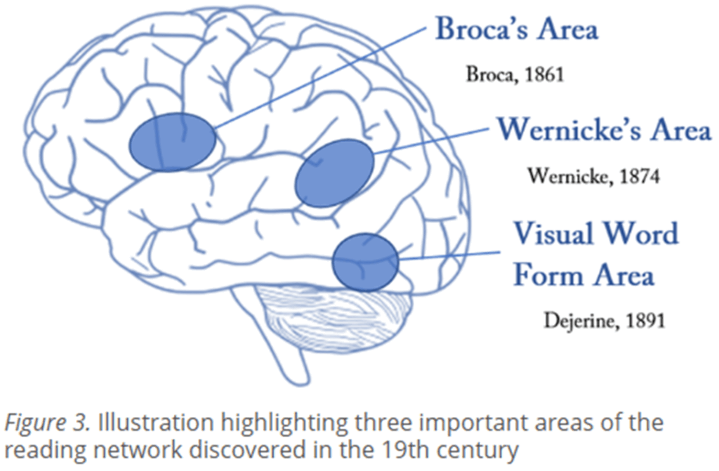
Two key brain regions involved in this theory are:
- Broca’s Area: Located in the left frontal lobe, this region is responsible for speech production and motor planning of language. Damage to Broca’s area can lead to slow, effortful speech with good comprehension (Broca’s aphasia).
- Wernicke’s Area: Found in the left temporal lobe, Wernicke’s area is responsible for language comprehension. Damage to this area can cause fluent but nonsensical speech and poor understanding (Wernicke’s aphasia).
The cerebral dominance theory of stuttering (Orton-Travis Theory, 1931)
In the 1920s, it was reported that many people who stutter were left-handed or ambidextrous.
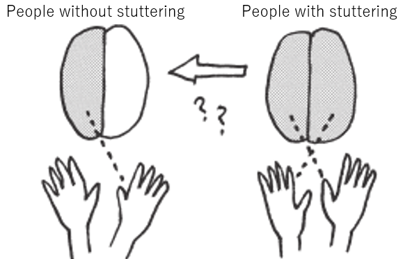
According to the cerebral dominance theory, if neither hemisphere developed dominance for language, or if both tried to control speech simultaneously, it could lead to confused neural signals and disorganized speech output—such as stuttering. This idea linked atypical handedness (e.g., left-handedness or ambidexterity) to speech difficulties.
In people who stutter, both cerebral hemispheres are thought to be equally involved in language processing.
One hypothesis suggests that stuttering occurs because the brain receives the same motor commands from both hemispheres at slightly different timings, leading to confusion about which command to follow.
Johnson rejected the cerebral dominance theory of stuttering (1942).
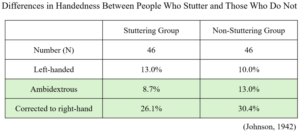
This table, based on Johnson’s 1942 study, compares handedness between people who stutter and those who do not. Both groups consisted of 46 individuals. The percentage of left-handed individuals was slightly higher in the stuttering group (13.0%) compared to the non-stuttering group (10.0%). In contrast, ambidextrous individuals were slightly more common in the non-stuttering group (13.0%) than in the stuttering group (8.7%). Interestingly, a similar proportion of individuals in both groups had been corrected to use their right hand, with 26.1% in the stuttering group and 30.4% in the non-stuttering group.
These small differences suggest that left-handedness, ambidexterity, and forced right-hand correction are not meaningfully related to the presence of stuttering. This evidence led Johnson to reject the cerebral dominance theory of stuttering, which had proposed that disruptions in hemispheric control, often inferred from handedness patterns, could cause stuttering.
Johnson’s diagnosogenic theory of stuttering (1940s)
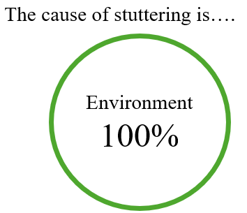
Stuttering is caused by anxious parents who react negatively to their child’s normal disfluencies.
Johnson developed indirect therapy to treat childhood stuttering.
The child doesn’t participate in the therapy. Instead, the parents change their behavior.
Evidence against diagnosogenic theory of stuttering
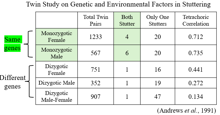
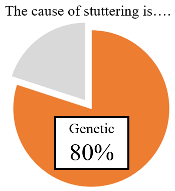
Among monozygotic twins, there are more cases where both individuals stutter compared to dizygotic twins. This suggests that genetic factors are involved in the cause of stuttering. A review of twin studies on stuttering indicates that approximately 80% of the cause of stuttering is related to genetic factors. (Frigerio-Domingues et al., 2017)
Examination of the Cerebral Dominance Theory (Brain Localization Analysis)
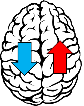
This diagram illustrates a modern interpretation of the cerebral dominance theory by showing differences in brain hemisphere function related to stuttering.
The left hemisphere (blue arrow pointing down) is shown as having impaired function, particularly in areas typically associated with speech production.
The right hemisphere (red arrow pointing up) shows increased compensatory activity, suggesting that it may be working harder to make up for the deficits in the left hemisphere.
In pediatric brain studies, increased activity in the right hemisphere is not observed in children who stutter. This suggests that the heightened right hemisphere activity often seen in adults may be a compensatory adaptation over time, rather than a root cause of stuttering.
Dual-Loop Model of Speech and Stuttering
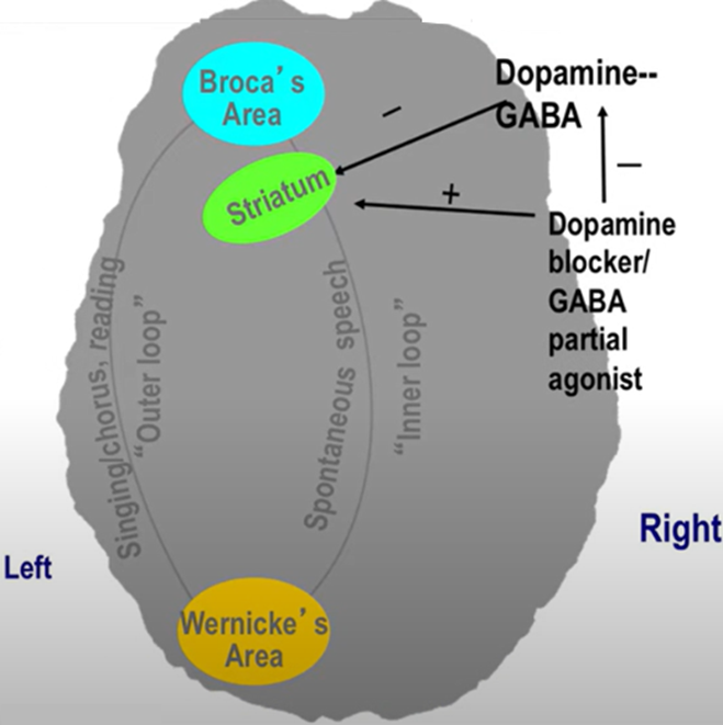
This figure illustrates the dual-loop model of speech processing and its relevance to stuttering. Functional brain imaging studies have shown that individuals who stutter exhibit reduced metabolism in both the cortical speech areas—Broca’s area and Wernicke’s area—and in the striatum, a part of the basal ganglia. These deficits are especially apparent during spontaneous speech, which relies on the “inner loop”, involving the striatum and modulated by dopamine. In contrast, tasks like singing or choral reading, which rely on the “outer loop”, bypass the striatal system and are often fluent in people who stutter.
The figure shows that dopaminergic overactivity disrupts the striatal function, impairing spontaneous speech.
Pharmacological interventions—dopamine blockers or GABA partial agonists—are thought to reduce this overactivity, thereby normalizing communication between the striatum and cortical speech areas. This model, based on Riley’s hypothesis, suggests that the outer loop remains functional in stuttering, while the inner loop, responsible for initiating speech, remains impaired due to striatal dysfunction.
(Ref.) Riley, G., Wu, J., & Maguire, G. (1997). PET scan evidence of parallel cerebral systems related to treatment effects. In W. Hulstijn, H. F. M. Peters, P. H. H. M. van Lieshout (Eds.), Speech production: Motor control, brain research, and fluency disorders (pp. 321–327). Amsterdam: Elsevier.
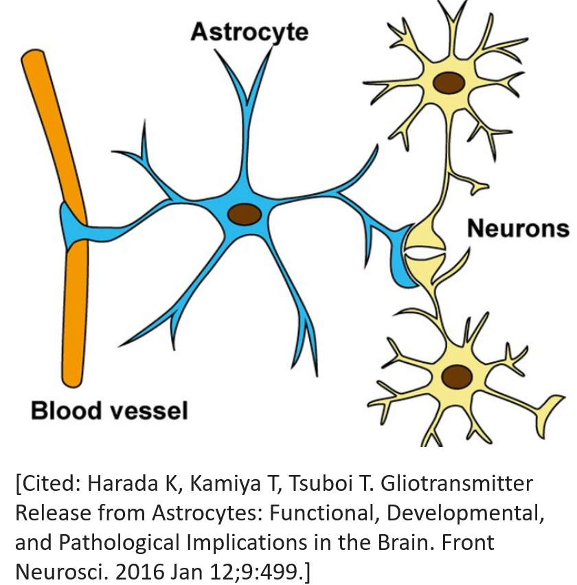
Astrocyte Dysfunction in the Neurobiology of Stuttering
Astrocyte Dysfunction in the Corpus Callosum: A Potential Cause of Stuttering in GNPTAB Mutant Mice
In the brains of GNPTAB mutant mice, there were no significant differences in neurons compared to controls; however, a marked reduction of astrocytes was specifically observed in the corpus callosum, the brain structure responsible for interhemispheric communication. This finding suggests that stuttering seen in these mice may arise from impaired astrocyte rather than from neuronal abnormalities[1].
Astrocytes are star-shaped cells in the brain that help neurons (the main brain cells) work properly. They don’t send messages like neurons do, but they have many important jobs:
- They support neurons. Astrocytes bring nutrients and oxygen to neurons to help them stay healthy and strong.
- They clean up waste. After neurons work, they make waste. Astrocytes help remove this waste to keep the brain clean.
- They help messages travel. Neurons send signals to each other. Astrocytes help control how strong or weak those signals are, so communication stays balanced.
- They protect the brain. Astrocytes help build the blood-brain barrier, which keeps harmful substances in the blood from entering the brain.
[1] Han TU, et al. Human GNPTAB stuttering mutations engineered into mice cause vocalization deficits and astrocyte pathology in the corpus callosum. Proc Natl Acad Sci U S A. 2019 Aug 27;116(35):17515-17524.
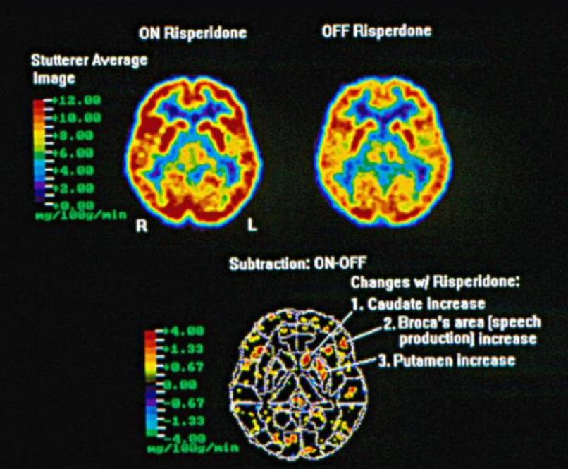
[2] Maguire GA, Yoo BR, SheikhBahaei S. Investigation of Risperidone Treatment Associated With Enhanced Brain Activity in Patients Who Stutter. Front Neurosci. 2021 Feb 12;15:598949.
Astrocytes may be involved in the brain function of individuals who stutter.
This study showed that people who stutter had increased brain activity in the striatum (caudate and putamen) and Broca’s area after taking the medication risperidone for six weeks.[2] These areas are involved in speech and motor control. The results support earlier findings that abnormal dopamine activity and reduced metabolism in the basal ganglia are central to stuttering.
Importantly, the study also suggests that astrocytes, a type of brain support cell, may be involved in the brain changes seen in stuttering. Astrocytes in the striatum and Broca’s area may become more active after risperidone treatment. Astrocytes can influence how brain signals are processed and may help regulate dopamine, a key brain chemical involved in movement and speech.
Because astrocytes have dopamine receptors and can be affected by medications like risperidone, the improved brain activity seen in this study could partly reflect increased astrocyte function.
These findings point to a new direction for research: studying how astrocytes and dopamine may contribute to stuttering, and how medications might help by restoring astrocyte function.
[2] Maguire GA, Yoo BR, SheikhBahaei S. Investigation of Risperidone Treatment Associated With Enhanced Brain Activity in Patients Who Stutter. Front Neurosci. 2021 Feb 12;15:598949.